Cells have caught the attention of electrical-engineering and neuroscience researchers recently. One team has been investigating an efficient, inexpensive way to count white blood cells, and another has focused on astrocytes, star-shaped brain cells believed to play a role in neurological disorders like Lou Gehrig’s, Alzheimer’s, and Huntington’s disease.
First, Hoseon Lee, an assistant professor of electrical engineering in the Southern Polytechnic College of Engineering and Engineering Technology at Kennesaw State University, has been researching ways for patients to monitor their white-blood-cell count without relying on flow cytometry, which generally requires bulky, expensive equipment and frequent, inconvenient trips to a hospital.
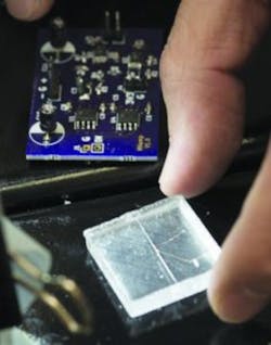
Courtesy of David Caselli, Kennesaw State University
“Flow cytometry equipment can perform lots of different functions: it can sort the cells, count them, and do other things that aren’t entirely necessary for every patient,” Lee said.1 “If we can focus on one thing—counting cells—we can make something smaller and more affordable for both the patient and the provider.”
Lee and six students have created a prototype (Figure 1) that uses a coil wrapped around a 100-μm-wide channel—large enough for two to three blood cells to pass through at a time. The channel is suspended in a small block of silicone gel, and the coil leads to a circuit board built by electrical engineering students to receive input as cells pass through the channel. The prototype operates on two AAA
batteries and could fit into a handheld configuration.
To count cells, the team attaches a magnetic nanoparticle to each white blood cell by mixing the two in a vial. As magnetized cells pass through the coil, a voltage flux is detected and logged. Each spike in voltage signifies a cell passing through the coil, providing a readout of white-blood-cell levels. Though the team is still perfecting its overall design, Lee said it has filed for a patent, and hopes to publish its findings in a peer-reviewed journal.
The research is somewhat personal, Lee said. His sister has a low white-blood-cell count that keeps her on a strict diet and requires a lot of rest. She lives in Seoul, South Korea, where traffic impedes her ability to visit the hospital for regular testing.
“She hates making that trip,” Lee said. “It pushed me to think of ways I could help her receive the same kind of monitoring without leaving her home.”
Studying astrocytes
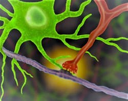
Second, UCLA neuroscientists have conducted research that could lead to a better understanding of astrocytes (Figure 2). In a recent paper, researchers describe a genetically targeted neuron-astrocyte proximity assay (NAPA). “NAPA provides a simple approach to measure astrocyte-synapse spatial interactions in a variety of experimental scenarios,” they write.2
Courtesy of UCLA/Khakh Lab
“We’re now able to see how astrocytes and synapses make physical contact and determine how these connections change in disorders like Alzheimer’s and Huntington’s disease,” said Baljit Khakh, a professor of physiology and neurobiology at the David Geffen School of Medicine at UCLA.3 “What we learn could open up
new strategies for treating those diseases—for example, by identifying cellular interactions that support normal brain function.”3
In the method created by Khakh’s team, different colors of light pass through a lens to magnify objects smaller than those viewable by earlier techniques. The new test allowed them to observe how interactions between synapses and astrocytes change over time, as well as during various diseases, in mouse models.
“This new tool makes possible experiments that we have been wanting to perform for many years,” added Khakh, a member of the UCLA Brain Research Institute. “For example, we can now observe how brain damage alters the way that astrocytes interact with neurons and develop strategies to address these changes.” EE
References


