Functional Connectivity MRI Helps Measure Brain Activity
Andre Jesmanowicz, associate professor at the Biophysics Research Institute of the Medical College of Wisconsin, is utilizing functional connectivity MRI (fcMRI) to enhance brain activity measurements. The technique reduces the time needed to acquire a single volumetric echo-planar imaging (EPI) data set. Whole brain studies require a full data set created using cross correlation of a seed voxel resting-state time course with other voxel time courses. The goal was to generate a whole brain scan in under two seconds.
Related Articles
- Digital Downconverter Provides Programmable Single & Dual Channels
- OpenVPX Helps Military Modules Mesh
- ReadyFlow Tools Enhance Cobalt User Experience
Multislice acquisition of 2D MR images usually processes one slice at a time. Scanning multiple slices at one time can accelerate the process, but this requires a more precise excitation process. This multislice excitation uses RF pulses with distinct phase tagging of each slice, which required a method to disentangle the information from overlapping slices. A Pentek Model 78621 dual-channel 800-MHz digital-to-analog converter (DAC) was used in the interpolating mode to create RF pulses with a 2-ns sampling rate and smooth stair-step-less modulation.
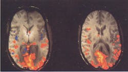
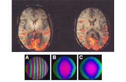
The synthesized RF pulses provide a coherent image (see the figure) compared to the first, conventionally obtained image. The scans were performed in conjunction with a GE Signa EXCITE 3 T MR Scanner. The accuracy of the DAC board provided a reference signal that compensated for the phase of off-resonance frequencies used to excite different slices.
The system obtains N slices for each of the n-number RF coil arrays. The first few slices are discarded and the remaining images are averaged to improve the signal-to-noise ratio (SNR). The result is N by n complex-valued reference images with good SNR.
MRIs have proven invaluable for diagnostics and research. Generating a more accurate MRI faster will help reduce the time patients need to spend in an MRI machine as well as provide more information to researchers.
About the Author
William G. Wong
Senior Content Director - Electronic Design and Microwaves & RF
I am Editor of Electronic Design focusing on embedded, software, and systems. As Senior Content Director, I also manage Microwaves & RF and I work with a great team of editors to provide engineers, programmers, developers and technical managers with interesting and useful articles and videos on a regular basis. Check out our free newsletters to see the latest content.
You can send press releases for new products for possible coverage on the website. I am also interested in receiving contributed articles for publishing on our website. Use our template and send to me along with a signed release form.
Check out my blog, AltEmbedded on Electronic Design, as well as his latest articles on this site that are listed below.
You can visit my social media via these links:
- AltEmbedded on Electronic Design
- Bill Wong on Facebook
- @AltEmbedded on Twitter
- Bill Wong on LinkedIn
I earned a Bachelor of Electrical Engineering at the Georgia Institute of Technology and a Masters in Computer Science from Rutgers University. I still do a bit of programming using everything from C and C++ to Rust and Ada/SPARK. I do a bit of PHP programming for Drupal websites. I have posted a few Drupal modules.
I still get a hand on software and electronic hardware. Some of this can be found on our Kit Close-Up video series. You can also see me on many of our TechXchange Talk videos. I am interested in a range of projects from robotics to artificial intelligence.


