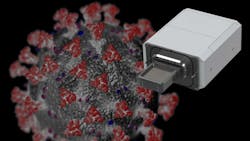
Cryogenic Electron Microscopy Method Assists in COVID-19 Vaccine Development (Download)
Advances in imaging technology continue to benefit society in disparate ways. For example, cryogenic electron microscopy (cryo-EM) played a fundamental role in the development of the COVID-19 vaccine. Combining cameras and detectors, sample-handling technology, automation, and software, cryo-EM is an easy-to-use method for high-quality data collection. It ultimately helped researchers identify the spike protein of SARS-CoV-2, the virus that causes COVID-19.
Cryo-EM involves flash-freezing a sample down to –180°C, leaving the protein particles suspended in ice. Sample sets are placed in a holder containing a grid that’s typically made of gold or carbon. Within the grid is foil with small holes or pockets. Material that’s imaged by the system is flash-frozen inside fluid contained in these holes.
